The ATP concentration in the cell is strictly regulated and is kept at a more or less stable level. The ATP determination serves to Evidence of an acquired, secondary mitochondriopathy. The investigation is not suitable for detecting an inherited, genetically determined functional disorder. A significant reduction in intracellular ATP is usually detected in the context of systemic inflammation.
Adenosine triphosphate, short ATP, is a molecule that provides energy as a storage substance (energy currency) in every cell of a living organism. This energy enables all work processes such as locomotion or metabolic processes in general. An ATP molecule contains three phosphate groups. To release energy, ATP is converted into ADP (adenosine diphosphate) by splitting off one of the three phosphate residues. The formation of ATP takes place in the mitochondria of the body's cells: By splitting sugar molecules, carbohydrates, fatty acids etc. from food, ATP, water and CO2 are formed with the help of oxygen as part of the respiratory chain:
Roughly speaking:
1 glucose +6 O2 +38 (ADP +P)
→ Respiratory chain citrate cycle →
6 CO2 + 6 H2O + 38 ATP
The ATP turnover per day is on average about half the human body mass, depending on the load.
Mitochondria
They are cell organelles and function as the cell's power plants in all living organisms. They provide energy in the form of ATP. And they can be damaged by environmental toxins (heavy metals, pesticides, insecticides, household poisons, cleaning agents, cosmetics), nanoparticles, antibiotics and other drugs, as well as stress and psychological pressure (mitochondriopathy). Neurological, metabolic, cardiac and oncological diseases are increasingly associated with a dysfunction of the mitochondria. Very energy-dependent organs/tissues are particularly affected by dysfunction: Brain, nerves, muscles.

When is ATP reduced in the cell?
The ATP concentration in the cell is strictly regulated and is kept at a more or less stable level, even if a lot is consumed at the moment, it is quickly regenerated. The ATP determination serves to Evidence of an acquired, secondary mitochondriopathy.
The investigation is not suitable for detecting an inherited, genetically determined functional disorder. A significant reduction in intracellular ATP is usually detected in the context of systemic inflammation. It often occurs together with laboratory diagnostic indications of immune activation (immune status, TNF-alpha, IP-10), and oxidative, nitrosative stress in the context of chronic inflammatory diseases such as chronic fatigue syndrome, cellular hypoxia, active viral infection, fibromyalgia, or chronic degenerative inflammatory processes. The determination of intracellular ATP therefore represents a important diagnostic parameters for the illustration of the current mitochondrial function.
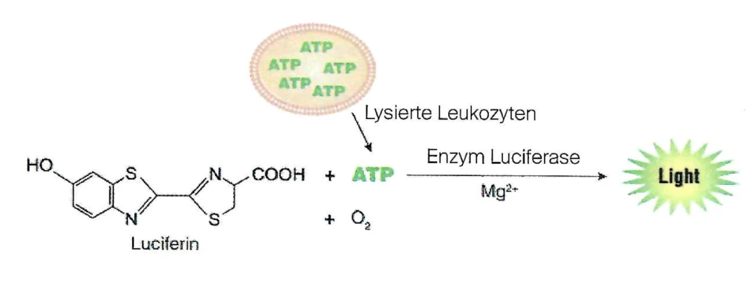
How is ATP measured?
Due to the high proportion of mitochondria in the granulocytes of the blood and their easy availability, these cells are ideal for the determination.
The leukocytes from heparinized whole blood are purified and the ATP is quantitatively detected in a defined number of cells after cell lysis using a specific chemiluminescence reaction (CLIA).
About Dr. med. Andreas Bernhardt:
Dr. Bernhardt is a specialist in general internal medicine with international training in endocrinology and Better Aging. He is a member of the Swiss Anti-Aging Society (SSAAMP) and the renowned Endocrine Society (Washington, D.C.). His focus is on bioidentical hormone therapy as part of a holistic longevity concept. As an expert on the German-speaking platform wechselweise.net he is committed to raising awareness in the DACH region about hormonal changes in men and women during the menopause - with the aim of promoting health and quality of life in the long term.
Literature:
Bell, C. J. et al: Luciferase expression for ATP imaging: application to cardiac myocytes. Methods in Cell Biology, 2007, 80: 341-352.
Crouch, S.P.M. et al. (1993) The use of ATP bioluminescence as a measure of cell proliferation and cytotoxicity. J. Immunol. Methods 160, 81-8.
Kangas, ,L. Grönroos, M. and Nieminen, AL. (1984) Bioluminescence of cellular ATP: Anew method for evaluating cytotoxic agents in vitro. Med. Biol. 62, 338-43.
Lundin, A. et al. (1986) Estimation of biomass in growing cell lines by adenosine triphosphate assay. Methods Enzymol. 13, 27-42.
Myhil, S. et al: Chronic fatigue syndrome and mitochondrial dysfunction. International Journal of Clinical and Experimental Medicine, 2009, :2 1-16.
Sevin, B.U. et al. (1988) Application of an ATP-bioluminescence assay in human tumor chemosensitivity testing. Gynecol Oncol. 31, 191-204.
Vernon, S. D. et al: Preliminary evidence of mitochondrial dysfunction associated with postinfective fatigue after acute infection with Epstein Barr virus. BMC Infectious Diseases, 2006, 6: 15.